Introduction
Our Departmental Research Centre for Medical Image Computing (RC-MIC) focuses on the development of new techniques for computation post-processing and medical image quantification, with the aim of developing computational methods to be used in everyday clinical diagnosis and clinically relevant research.
AI play an increasingly important role in the field of imaging research and clinical applications. Our Departmental CUHK lab of AI in radiology (CLAIR) focus on the use of AI for both acquisition and post-processing in medical imaging, with an aim to bring new technologies and optimize current technique to improve healthcare though multi disciplinary research involving.
To facilitate AI projects, the department has been equipped with high computational power GPU Cluster.
Some of our current work is focused on developing and applying various intelligent computational methodologies to facilitate the detection of imaging biomarkers for various neurological disorders.
On-going Research
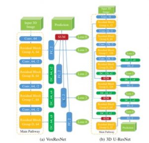
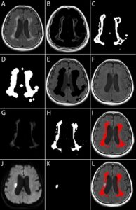
A Semi-Supervised Learning Approach to Automatic Segmentation of Major Manifestations of Small Vessel Disease in Multimodal Brain MR Images, General Research Fund, 01/01/2018 – 31/12/2020, HK$ 700,000
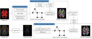
Description: Cerebral small vessel disease (SVD) is commonly presented in elderly and is considered to be associated with cognitive decline, functional loss, and increased stroke risk. Automatic identification of the major manifestations of SVD, i.e. recent small subcortical infarcts, white matter hyperintensities, lacunes, perivascular spaces, and microbleeds, would significantly improve the objectiveness of the SVD diagnosis and prognosis. In this project, a semi-supervised deep learning approach will be proposed to automatically identify the major manifestations of SVD in multimodal brain MR images. The novelties of the proposed method are summarized as follows. Firstly, a tailor-designed multi-modal input data structure is proposed to fit the architecture of 3D fully convolutional neural network. Secondly, in order to reduce the false positive and improve the detection accuracy of SVDs, an innovative deep learning structure is proposed with a nearly symmetric network structure which is ideal for handling the semantic information and localization simultaneously. In order to quantify the size of SVD lesions, a two-stage SVDs segmentation method composed of two different 3D FCNs is proposed and a dynamic weights of loss scheme to address the unbalanced-class problem. Thirdly, a new semi-supervised deep learning approach is proposed to train both the annotated and unannotated medical images. To assess the accuracy of proposed deep learning approach quantitatively by comparing with ground-truth. The application of this new method will be demonstrated in clinical researches, i.e., exploring the relationship between lesions and symptom of SVD and using as quantitative biomarkers in drug trials.
Development of a Machine-learning Approach Computational Vascular Diagnoses Platform of Fractional Flow Reserve from Multimodality Medical Imaging. Innovation and Technology Support Programme, Innovation and Technology Support Programme, 1/9/2017-31/8/2019, HK$ 5,000,000
Description: The luminal stenosis and ischemia caused by Atherosclerosis is the culprit leading to cardiovascular disease. Doctors can get a better understand of the clinical course on basis of patient specific hemodynamic data. Efficient and accurate estimation of vasculopathy is one of most critical parts for setting the therapeutic strategies. Currently, the hemodynamic results based on Computational Fluid Dynamics are largely dependent on the setting of the boundary condition. The reliability has a limitation as well as it is very time consuming and complicated which is difficult to clinical promotion. Thus, we would like to present a brand new computer platform for hemodynamic simulation based on machine-learning approach as an alternative to physics-based approaches. This platform could not only saves a great deal of time, but also makes the analyzing result more accurate and reliable. The machine-learning model is trained on a large database of synthetic vessel trees, followed by verification and validation against an existing physics-based model, as well as invasive measurements. With this platform, we would learn the behavior of complex physiological models with proven capability of predicting invasive FFR, using purely anatomical information available in the clinical routine.
Past Research
Seeing the Truth of Human Body – The Next-Generation Glasses-Free 3D Visualization Suite for Medical Images
Principal Investigator – Professor Shi Lin
Ref No: ITS/293/14FP, Innovation and Technology Support Programme, 01/10/2015 – 30/09/2017, HKD 1,604,000
Detecting distinctive neuro-connectivity biomarkers for patients with vascular cognitive impairment–An EEG-informed fMRI approach
Principal Investigator – Professor Shi Lin
General Research Fund, 01/01/2015 – 31/12/2017, HKD 908,730
A computer-assisted platform for BoneBridge hearing aid implantation surgery
Principal Investigator – Professor Shi Lin
Project Code: ITS/225/12, 01/09/2013 – 29/02/2015, HKD 999,800
Detection and Grading of Cam-type Femoroacetabular Impingement (FAI) from Radiological Signs Using Computerized Methods
Principal Investigator – Professor Shi Lin
General Research Fund, Project Code: 473012, 01/01/2013 – 31/12/2015, HKD 695,460
Algorithm Development for Knowledge based Spinal MRI Segmentation (基于知识的脊柱MRI影像分割算法研究)
Principal Investigator – Professor Shi Lin
National Natural Science Foundation of China (NSFC), Project Code: 81101111; 01/01/2012-31/12/2014, RMB 230,000
Mapping the contribution and strategic distribution patterns of neuroimaging features of small vessel disease in post-stroke cognitive impairment. Journal of Neurology, Neurosurgery & Psychiatry. (Accepted) (IF: 7.35)
Lin Shi, Lei Zhao, Fu Ki Yeung, Shun Yiu Wong, King To Chan, … Jill Abrigo, Winnie Chu, Vincent CT Mok
ABSTRACT
OBJECTIVES: Individual neuroimaging features of small vessel disease (SVD) have been reported to influence poststroke cognition. This study aimed to investigate the joint contribution and strategic distribution patterns of multiple types of SVD imaging features in poststroke cognitive impairment.
METHODS: We studied 145 first-ever ischaemic stroke patients with MRI and Montreal Cognitive Assessment (MoCA) examined at baseline. The local burdens of acute ischaemic lesion (AIL), white matter hyperintensity, lacune, enlarged perivascular space and cross-sectional atrophy were quantified and entered into support vector regression (SVR) models to associate with the global and domain scores of MoCA. The SVR models were optimised with feature selection through 10-fold cross-validations. The contribution of SVD features to MoCA scores was measured by the prediction accuracy in the corresponding SVR model after optimisation.
RESULTS: The combination of the neuroimaging features of SVD contributed much more to the MoCA deficits on top of AILs compared with individual SVD features, and the cognitive impact of different individual SVD features was generally similar. As identified by the optimal SVR models, the important SVD-affected regions were mainly located in the basal ganglia and white matter around it, although the specific regions varied for MoCA and its domains.
CONCLUSIONS: Multiple types of SVD neuroimaging features jointly had a significant impact on global and domain cognitive functionings after stroke on top of AILs. The map of strategic cognitive-relevant regions of SVD features may help clinicians to understand their complementary impact on poststroke cognition.
Automatic Segmentation of Acute Ischemic Stroke from DWI using 3D Fully Convolutional DenseNets. IEEE Transactions on Medical Imaging. (Accepted) (IF:3.94)
Rongzhao Zhang, Lei Zhao, Wutao Lou, Jill M Abrigo, Vincent CT Mok, Winnie CW Chu, Defeng Wang, Lin Shi*
ABSTRACT
Acute ischemic stroke is recognized as a common cerebral vascular disease in aging people. Accurate diagnosis and timely treatment can effectively improve the blood supply of the ischemic area and reduce the risk of disability or even death. Understanding the location and size of infarcts plays a critical role in the diagnosis decision. However, manual localization and quantification of stroke lesions are laborious and timeconsuming. In this paper, we propose a novel automatic method to segment acute ischemic stroke from diffusion weighted images (DWI) using deep 3D convolutional neural networks (CNNs). Our method can efficiently utilize 3D contextual information and automatically learn very discriminative features in an end-to-end and data-driven way. To relieve the difficulty of training very deep 3D CNN, we equip our network with dense connectivity to enable the unimpeded propagation of information and gradients throughout the network. We train our model with Dice objective function to combat the severe class imbalance problem in data. A DWI dataset containing 242 subjects (90 for training, 62 for validation and 90 for testing) with various types of acute ischemic stroke was constructed to evaluate our method. Our model achieved high performance on various metrics (Dice similarity coefficient: 79.13%, lesion-wise precision: 92.67%, lesion-wise F1 score: 89.25%), outperforming other state-of-the-art CNN methods by a large margin. We also evaluated the model on ISLES2015- SSIS dataset and achieved very competitive performance, which further demonstrated its generalization capacity. The proposed method is fast and accurate, demonstrating a good potential in clinical routines.
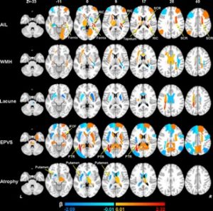
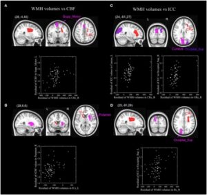
The Spatial Associations of Cerebral Blood Flow and Spontaneous Brain Activities with White Matter Hyperintensities – An Exploratory Study Using Multimodal MRI. Frontiers in Neurology, 2017; 8:593. (IF: 3.552)
Lin Shi, Xinyuan Miao, Wutao Lou, Kai Liu, Jill Abrigo, Adrian Wong, Winnie C.W. Chu, Defeng Wang, Vincent Chung Tong Mok
ABSTRACT
White matter hyperintensities (WMHs) have been reported to be correlated with functional brain changes, but the association of the specific WMHs distribution pattern with regional functional changes remains uncertain. The aim of this study is to explore the possible spatial correlation of WMH with changes in cerebral blood flow (CBF) and spontaneous brain activities in elderly using a novel approach. The WMHs, CBF, and spontaneous brain activities measured by intrinsic connectivity contrast (ICC), were quantified using multimodal magnetic resonance imaging for 69 elderly subjects. Such approach enables us to expand our search for newly identified correlated areas by drawing strengths of different modes and provides a means for triangulation as well as complementary insights. The results showed significant positive correlations between WMH volumes in the right superior corona radiata and CBF in the left supplementary motor area, as well as between WMH volumes in left anterior limb internal capsule and CBF in the right putamen. Significant correlations of regional WMH volumes and ICC were also detected between the right anterior corona radiata and the left cuneus, and the right superior occipital cortex, as well as between the right superior corona radiata and the left superior occipital cortex. These findings may suggest a regional compensatory functional enhancement accounting for the maintenance of cognitively normal status, which can be supported by the widely observed phenomenon that mild to moderate WMH load could have little effect on global cognitive performance.
Supervoxel-based Statistical Analysis of Diffusion Tensor Imaging in Schizotypal Personality Disorder. NeuroImage, 2017; 163:368-378. (IF: 835)
Teng Zhang, Defeng Wang, Qing Zhang, Jianlin Wu, Jian Lv, Lin Shi*
ABSTRACT
To study white matter changes in schizotypal personality disorder (SPD), we developed a new statistical analysis method based on supervoxels for diffusion tensor imaging. Twenty patients with SPD and eighteen healthy controls were recruited from a pool of 3000 first-year university undergraduates, and underwent MRI using a 3T scanner. Diffusion tensors were first normalized into ICBM-152 space followed by a supervoxel segmentation based on graph clustering to segment white matter tensors into diffusion homogeneous supervoxels. Fractional anisotropy (FA) values in supervoxels were compared between SPD and healthy controls using permutation test. Suprathreshold cluster size test was used to correct multiple comparison. At last, fibers with significant differences were extracted from supervoxel clusters with significance level P < 0.05. Results showed that FA values in genu of corpus callosum were significantly reduced (P = 0.012) in patients with SPD (FA = 0.565) compared with healthy controls (FA = 0.593). In summary, this study proposed a novel supervoxel segmentation method for diffusion tensor imaging using graph-based clustering, and extended permutation test and suprathreshold cluster size test to supervoxels for detection of white matter changes.
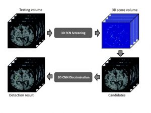
Automatic Detection of Cerebral Microbleeds from MR Images via 3D Convolutional Neural Networks. IEEE Trans. on Medical Imaging 2016;35(5):1182-95. (IF: 3.756)
Qi Dou, Hao Chen, Lequan Yu, Lei Zhao, Jing Qin, Defeng Wang, Vincent CT Mok, Lin Shi*, Pheng-Ann Heng
ABSTRACT
Cerebral microbleeds (CMBs) are small haemorrhages nearby blood vessels. They have been recognized as important diagnostic biomarkers for many cerebrovascular diseases and cognitive dysfunctions. In current clinical routine, CMBs are manually labelled by radiologists but this procedure is laborious, time-consuming, and error prone. In this paper, we propose a novel automatic method to detect CMBs from magnetic resonance (MR) images by exploiting the 3D convolutional neural network (CNN). Compared with previous methods that employed either low-level hand-crafted descriptors or 2D CNNs, our method can take full advantage of spatial contextual information in MR volumes to extract more representative high-level features for CMBs, and hence achieve a much better detection accuracy. To further improve the detection performance while reducing the computational cost, we propose a cascaded framework under 3D CNNs for the task of CMB detection. We first exploit a 3D fully convolutional network (FCN) strategy to retrieve the candidates with high probabilities of being CMBs, and then apply a well-trained 3D CNN discrimination model to distinguish CMBs from hard mimics. Compared with traditional sliding window strategy, the proposed 3D FCN strategy can remove massive redundant computations and dramatically speed up the detectionprocess. We constructed a large dataset with 320 volumetric MR scans and performed extensive experiments to validate the proposed method, which achieved a high sensitivity of 93.16% with an average number of 2.74 false positives per subject, outperforming previous methods using low-level descriptors or 2D CNNs by a significant margin. The proposed method, in principle, can be adapted to other biomarker detection tasks from volumetric medical data.
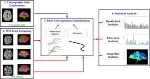
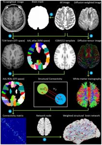
Tractography Atlas-based Spatial Statistics: Statistical Analysis of Diffusion Tensor Image along Fiber Pathways, NeuroImage, 2016;125:301-10. (IF: 5.835)
Defeng Wang, Yishan Luo, Vincent Mok, Winnie CW Chu, Lin Shi*
ABSTRACT
The quantitative analysis of diffusion tensor image (DTI) data has attracted increasing attention in recent decades for studying white matter (WM) integrity and development. Among the current DTI analysis methods, tract-based spatial statistics (TBSS), as a pioneering approach for the voxelwise analysis of DTI data, has gained a lot of popularity due to its user-friendly framework. However, in recent years, the reliability and interpretability of TBSS have been challenged by several works, and several improvements over the original TBSS pipeline have been suggested. In this paper, we propose a new DTI statistical analysis method, named tractography atlas-based spatial statistics (TABSS). It doesn’t rely on the accurate alignment of fractional anisotropy (FA) images for population analysis and gets rid of the skeletonization procedures of TBSS, which have been indicated as the major sources of error. Furthermore, TABSS improves the interpretability of results by directly reporting the resulting statistics on WM tracts, waiving the need of a WM atlas in the interpretation of the results. The feasibility of TABSS was evaluated in an example study to show age-related FA alternation pattern of healthy human brain. Through this preliminary study, it is validated that TABSS can provide detailed statistical results in a comprehensive and easy-to-understand way.
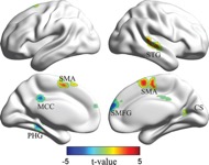
Changes of Cerebral Perfusion and Functional Brain Network Organization in Patients with Mild Cognitive Impairment, Journal of Alzheimer’s Disease, 2016; 54(1):397-409
Wutao Lou, Lin Shi*, Adrian Wong, Winnie C.W. Chu, Vincent C.T. Mok, and Defeng Wang, C
ABSTRACT
Disruptions of the functional brain network and cerebral blood flow (CBF) have been revealed in patients with mild cognitive impairment(MCI). However, the neurophysiological mechanism of hypoperfusion as well as the reorganization of the intrinsic whole brain network due to the neuropathology of MCI are still unclear. In this study, we aimed to investigate the changes of CBF and the whole brain networkorganization in MCI by using a multimodal MRI approach. Resting state ASL MRI and BOLD MRI were used to evaluate disruptions of CBF and underlying functional connectivity in 27 patients with MCI and 35 cognitive normal controls (NC). The eigenvector centrality mapping (ECM) was used to assess the whole brain network reorganization in MCI, and a seed-based ECM approach was proposed to reveal the contributions of the whole brain network on the ECM alterations. Significantly decreased perfusion in the posterior parietal cortex as well as its connectivity within the default mode network and occipital cortex were found in the MCI group compared to the NC group. The ECM analysis revealed decreased EC in the middle cingulate cortex, parahippocampal gyrus, medial frontal gyrus, and increased EC in the right calcarine sulcus, superior temporal gyrus, and supplementary motor area in the MCI group. The results of this study indicate that there are deficits in cerebral blood flow and functional connectivity in the default mode network, and that sensory-processing networks might play a compensatory role to make up for the decreased connections in MCI.
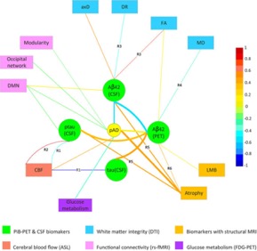
Mapping the Relationship of Contributing Factors for Preclinical Alzheimer’s Disease. Scientific Reports, Article number: 11259 (2015)
Lin Shi, Lei Zhao, Adrian Wong, Defeng Wang, and Vincent Mok
ABSTRACT
While detecting and validating correlations among the contributing factors to the preclinical phase of Alzheimer’s disease (pAD) has been a focus, a potent meta-analysis method to integrate current findings is essential. The entity-relationship diagram with nodes as entities and edges as relationships is a graphical representation that summarizes the relationships among multiple factors in an intuitive manner. Based on this concept, a new meta-analysis approach with this type of diagram is proposed to summarize research about contributing factors of pAD and their interactions. To utilize the information for enriched visualization, width and color of the edges are encoded with reporting times, number of pAD subjects, correlation coefficient, and study design (cross-sectional or longitudinal). The proposed Probabilistic Entity-Relationship Diagram (PERD) demonstrated its effectiveness in this research for studying pAD. Another kind of diagram with occurrence order for some factors was also proposed to provide sequential information of the factors. In addition, PERD could potentially develop into an online application named PERD-online, which would help researchers to pool findings on the same relationships and guide further tests to validate uncertain relationships in PERD. PERD as a generic graphical meta-analysis tool can also be applied in studying other multifactorial diseases.
Construction of brain atlases based on a multi-center MRI dataset of 2020 Chinese adults, Scientific Reports (2015)
Peipeng Liang, Lin Shi (equal contributor as the first author), Nan Chen, Yishan Luo, Xing Wang, Kai Liu, Vincent Mok, Winnie CW Chu, Defeng Wang, Kuncheng Li
ABSTRACT
Despite the known morphological differences (e.g., brain shape and size) in the brains of populations of different origins (e.g., age and race), the Chinese brain atlas is less studied. In the current study, we developed a statistical brain atlas based on a multi-center high quality magnetic resonance imaging (MRI) dataset of 2020 Chinese adults (18-76 years old). We constructed 12 Chinese brain atlas from the age 20 year to the age 75 at a 5 years interval. New Chinese brain standard space, coordinates, and brain area labels were further defined. The new Chinese brain atlas was validated in brain registration and segmentation. It was found that, as contrast to the MNI152 template, the proposed Chinese atlas showed higher accuracy in hippocampus segmentation and relatively smaller shape deformations during registration. These results indicate that a population-specific time varying brain atlas may be more appropriate for studies involving Chinese populations.
Abnormal Organization of White Matter Network in Patients with noDementia after Ischemic Stroke. Plos One, 2013, 8(12):e81388
Lin Shi, Defeng Wang, Winnie CW Chu, Shangping Liu, YunyunXiong, Yilong Wang, Yongjun Wang, Lawrence KS Wong, Vincent CT Mok
ABSTRACT
Structural changes after ischemic stroke could affect information communication extensively in the brain network. It is likely that the defects in the white matter (WM) network play a key role in information interchange. In this study, we used graph theoretical analysis to examine potential organization alteration in the WM network architecture derived from diffusion tensor images from subjects with no dementia and experienced stroke in the past 5.4-14.8 months (N = 47, Mini-Mental Screening Examination, MMSE range 18-30), compared with a normal control group with 44 age and gender-matched healthy volunteers (MMSE range 26-30). Region-wise connectivity was derived from fiber connection density of 90 different cortical and subcortical parcellations across the whole brain. Both normal controls and patients with chronic stroke exhibited efficient small-world properties in their WM structural networks. Compared with normal controls, topological efficiency was basically unaltered in the patients with chronic stroke, as reflected by unchanged local and global clustering coefficient, characteristic path length, and regional efficiency. No significant difference in hub distribution was found between normal control and patient groups. Patientswith chronic stroke, however, were found to have reduced betweenness centrality and predominantly located in the orbitofrontal cortex, whereas increased betweenness centrality and vulnerability were observed in parietal-occipital cortex. The National Institutes of Health Stroke Scale (NIHSS) score of patient is correlated with the betweenness centrality of right pallidum and local clustering coefficient of left superior occipital gyrus. Our findings suggest that patients with chronic stroke still exhibit efficient small-world organization and unaltered topological efficiency, with altered topology at orbitofrontal cortex and parietal-occipital cortex in the overall structural network. Findings from this study could help in understanding the mechanism of cognitive impairment and functional compensation occurred in patients with chronic stroke.
Intensity and Sulci Landmark Combined Brain Atlas Construction for Chinese Pediatric Population, Human Brain Mapping, 35(8):3880-92 (2014)
Yishan LUO, Lin SHI, Jian WENG, Hongjian HE, Winnie CW CHU, Feiyan CHEN, Defeng WANG
ABSTRACT
Constructing an atlas from a population of brain images is of vital importance to medical image analysis. Especially in neuroscience study, creating a brain atlas is useful for intra- and inter-population comparison. Research on brain atlas construction has attracted great attention in recent years, but the research on pediatric population is still limited, mainly due to the limited availability and the relatively low quality of pediatric magnetic resonance brain images. This article is targeted at creating a high quality representative brain atlas for Chinese pediatricpopulation. To achieve this goal, we have designed a set of preprocessing procedures to improve the image quality and developed an intensity and sulci landmark combined groupwise registration method to align the population of images for atlas construction. As demonstrated in experiments, the newly constructed atlas can better represent the size and shape of brains of Chinese pediatric population, and show better performance in Chinese pediatric brain image analysis compared with other standard atlases.
Automated Quantification of White Matter Lesion in Magnetic Resonance Imaging of Patients with Acute Infarction. Journal of Neuroscience Methods. 213(1), Pages 138–146 (2013)
Lin Shi, Defeng Wang, Shangping Liu, Yuehua Pu, Yilong Wang, Winnie CW Chu, Anil T Ahuja, Yongjun Wang
ABSTRACT
PURPOSE: It has been reported that increased white matter lesions (WML) is one of the risk factors for stroke. To quantify WML objectively with the presence of acute infarcts, we proposed an automated segmentation scheme to locate WMLs in combined T1-weighted MRI, fluid attenuation inversion recovery (FLAIR) and diffusion weighted imaging (DWI).
MATERIALS AND METHODS: The proposed method detects WMLs by a coarse-to-fine mathematical morphology method. It has been evaluated quantitatively and qualitatively using voxel-based, volume-based, score-based, and atlas-based approaches on MRI data of 91 subjects with acute infarction.
RESULT: The proposed WML detection algorithm yields average sensitivity, positive predictive value and similarity index of 0.803, 0.818, and 0.836, respectively. Experimental results demonstrated that the segmentation from the proposed method is in high agreement with that from manual segmentation (intraclass correlation coefficient=0.9892), and with a good correlation with visual scores (R=0.8442, p<0.0001).
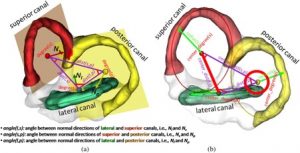
MRI Segmentation and Morphoanatomy Analysis of the Vestibular System in Adolescent Idiopathic Scoliosis, NeuroImage, 54 Suppl 1:S180-8. (2011)
Lin Shi, Defeng Wang, Winnie C.W. Chu, Tien-Tsin Wong, Geoffrey R. Burwell, and Jack C.Y. Cheng, Pheng Ann Heng
ABSTRACT
The vestibular system is the sensory organ responsible for perceiving head rotational movements and maintaining postural balance of human body. The objectives of this study are to propose an innovative computational technique capable of automatically segmenting the vestibularsystem and to analyze its geometrical features from high resolution T2-weighted MR images. In this study, the proposed technique was used to test the hypothesis that the morphoanatomy of vestibular system in adolescent idiopathic scoliosis (AIS) patients is different from healthy control subjects. The findings could contribute significantly to the understanding of the etiopathogenesis of AIS. The segmentation pipeline consisted of extraction of region of interest, image pre-processing, K-means clustering, and surface smoothing. The geometry of this high-genus labyrinth structure was analyzed through automatic partition into genus-0 units and approximation using the best-fit circle and plane for each unit. The metrics of the best-fit planes and circles were taken as shape measures. The proposed technique was applied on a cohort of 20 right-thoracic AIS patients (mean age 14.7 years old) and 20 age-matched healthy girls. The intermediate results were validated by subjective scoring. The result showed that the distance between centers of lateral and superior canals and the angle with vertex at the center of posterior canal were significantly smaller in AIS than in healthy controls in the left-side vestibular system with p=0.0264 and p=0.0200 respectively, but not in the right-side counterparts. The detected morphoanatomical changes are likely to be associated with subclinical postural, vestibular and proprioceptive dysfunctions reported frequently in AIS. This study has demonstrated that the proposed method could be applied in MRI-based morphoanatomy studies of vestibular system clinically.